Digitizing bioprocesses.in
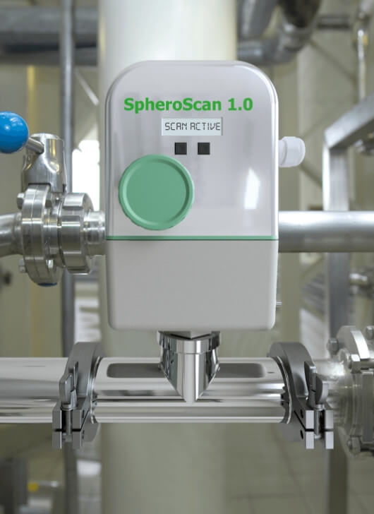
We bring advanced pathogen detection in pocket-size instruments directly to the production site, enabling seamless process monitoring and control.
Today’s bio-analytical methods are confined to remote laboratories. Our sensor technology uses light-emitting µBeads as a messenger to provide in-line, real-time pathogen detection inside liquid products along the entire process chain. Our µBeads work in vitro and in situ and can be tailored to be highly target-specific.
Thus, we enhance food safety and help improve production processes.
Basic Technology
Biosensors
Conventional biosensors (Figure 1 left) typically guide the analyte under study through a microchannel, in which one wall is functionalized by means of specific capture molecules, so that only wanted targets may selectively bind. The binding reactions are then converted into electrical signals via some physical transducer mechanism.
Drawbacks of conventional biosensors:
1. Laminar flow impedes the movement of targets towards the sensor area
2. Unspecific binding to the sensor area is not effectively flushed away
3. The cost of these sensors is high, and they are difficult to regenerate or replace
Biosensors based on Fluorescent Resonator Signature (FRS) technology, on the other hand, use freely moving sensor particles. Also here, the biospecific interface consists of specific capture molecules, but now these are grafted onto the surface of the microscopic sensor particles. Since the particles are freely floating, the entire analyte volume may be scanned for target molecules within short, thus greatly improving the reliability of the measurement. By means of our proprietary approach, binding events are read out optically and thus contact-less, and subsequently converted into electrical signals.
Disruptive characteristics of Fluorescence Resonator Signature (FRS):
1. High number of freely floating µBeads scan entire analyte volume
2. Spherical sensing surface avoids non-specific binding as well as specific re-binding
3. Optical technology allows for contact-less read-out of each individual µBead sensor
Figure 1: Comparison of a common biosensor (left) with biosensor based on Fluorescent Resonator Signature (right).
µBeads as free-floating sensor particles
SpheroScan® makes use of a novel approach to label-free bacterial and fungal detection. It is based on fluorescent, specifically functionalized polymer µBeads, which bind to their target organism while freely floating in the analyte.
Figure 2: left: illustration of µBead functionalization and target binding; right: SEM micrograph of 10 µm µBead decorated with specifically bound legionella
Depending on the respective application, size and optical properties of the µBeads are chosen for best performance even in complex media, while their surface can be functionalized with different capture molecules, such as peptides, aptamers, or antibodies. Quality control after each step of preparation warrants for reproducible and reliable performance of the overall system.
Figure 3: Examples for surface functionalization depending on sensing application
Remote read-out of the µBeads
Sensor read-out is performed remotely by optical means, i.e., fluorescent excitation of the organic dye embedded into the polymer beads and recording of their fluorescent emission (Fig. 4 left). As illustrated in Fig. 4, right, this fluorescent emission undergoes characteristic changes upon binding of the target species on the sensor surface (Fluorescent Resonator Signature, FRS). A numeric algorithm based on Mie theory is then applied to the resultant fluorescence spectra for quantification of the bound target. Since the sensors are microscopic particles, their number can be made sufficiently high to allow for reliable statistics on the concentration of the targeted microbe.
Figure 4: Schematic of Fluorescent Resonator Signature (FRS) Sensing: (left) optical set-up for excitation and readout of FRS of the freely floating sensor beads; (right) illustration of the change in FRS by specific binding of some targeted microbe to the µBead surface.
Results
The algorithm first calculates the µBead radius and the layer thickness (see Figure 5) of the connected target on the surface. The signal can then be converted to the concentration of the target in the medium (e.g. CFU, ppm or µg/l) using a stored calibration.
Figure 5: Basic results of FRS proprietary Algorithm: bead size distribution (left) and surface load upon protein adsorption (right)
Fields Of Application
A sensor right where quality matters the most.
Fermentation, Production
Real-time, inline pathogen detection and yeast health monitoring in production facilities for food or alcohol, e.g. Bio-Ethanol, results in an optimized process and leads to increased efficiency.
Agriculture & Food Processing
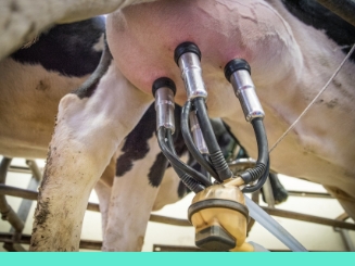
Real-time, inline pathogen detection in farm, food, and beverage production to reduce waste and increase quality.
Clean Water, Public Health
Pathogen detection for quality control of fresh water and disease control in waste water.
Healthcare
Diagnostics
in vivo or in vitro
coming soon…
FluIDect GmbH
If you have any questions, please contact us.
Address
Winzerlaer Str. 2, 07745 Jena, Germany
Call Us
+49 (0) 3641 / 55 41 380
Email Us
info@fluidect.com